Multimedia embryos
Now that scientific publishing has largely moved online, we are used to journal articles surrounded by supplementary material. Not just press releases and editorials, but also data files, podcasts and videos – without them no self-respecting breakthrough is complete. The scientific article, for over a century the gold standard of publication in the sciences, has become a multimedia spectacle, accessible at our desks, on screen. By contrast, runs of old journals, not to mention digitised backfiles, can seem a bit thin: just text and illustrations on the page. Yet this impression can mislead. It is an artefact of our assumptions and what libraries keep. Publishing a hundred years ago could be as richly complex and – this is the real difference – reading far more intensely physical than today.
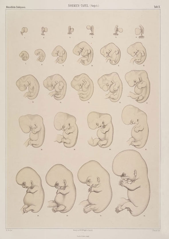
Antiquarian booksellers still list 'three parts plus atlas', but the founding work of modern human embryology was once much more. Colleagues in the 1880s recognized the Anatomy of Human Embryos by the Leipzig anatomist Wilhelm His as fully three 'publication series': two folio atlases, three instalments of text (part 1 and part 3) plus related articles, and two 'inseparable' sets of wax models.

The late nineteenth century was a great age of print. In academic publishing, especially in and around the German universities, one journal, textbook and handbook was founded after another. And yet, in fields such as human embryology, scientists learned at the same time to 'publish' in wax as well as print. Mixing media, their works are full of interactions between two dimensions and three.
Embryology, which told how complex adult bodies develop from apparently simple beginnings, was a major science of human origins. But its objects – tiny, intricate, translucent and often very scarce – presented a huge challenge. Human embryos, hidden in pregnant bodies, were especially difficult to study. Most material came from abortions, spontaneous or induced, and no specimens from the first two to three weeks were known. Human embryos also attracted special interest in the post-Darwinian debates over the theory of evolution. Evolutionists claimed them as proxies for absent fossils.
Critical of the leading Darwinists, and of descriptions by the clinicians who had direct access to specimens, from the late 1870s Wilhelm His reformed the field. He analysed an unprecedented number of embryos from the third week to the eighth month. This involved organizing a network of collectors, who framed as embryos blood clots that aborting women had usually interpreted in very different terms and handed them over to him. He dissected the embryos out of the egg membranes, placed them in developmental order and selected the most normal-looking as 'norms'. Above all, he analysed them with demanding new techniques.
His had invented the first microtome, or mounted knife, capable of producing serial slices, or sections, through an embryo. This gave much more detailed access to microscopic internal structures, but at the risk of fragmenting understanding. Embryologists already used models extensively in teaching, to help medical students grasp the complex changes in shape. His now insisted that researchers not only section even such 'treasures' as early human embryos, but also make models that would 'give' them 'body' again. At first, he worked freehand with spatulas and spoons. Later he adopted a method of cutting the highly magnified outlines of the structures of interest from wax plates and stacking these up. (Find out more about publishing in wax and print and the wax-plate modelling technique.)

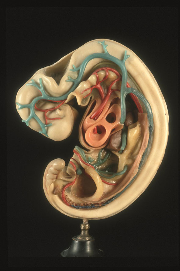
His gave his publisher the manuscripts for the Anatomy of Human Embryos. He sent the original models to the modeller Friedrich Ziegler, who reproduced them in coloured wax. One series shows whole embryos, another 'dissects' them; still more focus on hearts and brains.
Ziegler used the language of print, writing of 'authors' and 'proofs'. These were the first set of models, to which the scientist could ask for corrections before allowing them to go out under his or her name. But the business was very different, with new sets made to order and distribution in special crates. The time-consuming, fiddly, skilled labour made the models expensive. But they were so indispensable to understanding His's work, and to teaching it, that people put up with the extra trouble and cost to make them available in all universities and colleges where embryology was taught. The Anatomy of Human Embryos was read with models on the bench.
Some scientists still disparaged models as suitable only for teaching, or even as belonging in popular anatomy museums, alongside obstetric operations and syphilitic penises. The anatomists who followed His in human and comparative vertebrate embryology disagreed. They sectioned embryos and reconstructed highly specialized models which their articles or monographs described and depicted and Ziegler reproduced. The anatomists, used to learning by cutting, especially valued the physicality of the interaction.
These extraordinary objects went out of fashion long ago, though some were used in teaching until recently and many are still to be found in older institutes and museums. They are the solid, heavy, static ancestors of the light, bright, dynamic forms that now rotate in the flat '3-D' space of our screens. They are also the original 'supplementary material'. Readers could not call it up with a click and it was certainly not available on open access, but in part for that very reason the experience could be more profound: more physical and more unusual. As we look through old books and journals online, we should remember that some were never intended to stand alone, but were designed as components of multimedia displays.
Nick Hopwood (HPS, Cambridge)
Further reading
There is much more about embryos, books and models in the online exhibition on Making Visible Embryos. For the work of Wilhelm His, see especially 'Remodelling'.
Nick Hopwood, 'Model politics', The Lancet 372 (6 December 2008), 1946–7
Soraya de Chadarevian and Nick Hopwood (eds), Models: The Third Dimension of Science (Stanford: Stanford University Press, 2004)
Nick Hopwood, Embryos in Wax: Models from the Ziegler Studio, with a Reprint of 'Embryological Wax Models' by Friedrich Ziegler (Whipple Museum of the History of Science, University of Cambridge; Institute of the History of Medicine, University of Bern, 2002)
Nick Hopwood, 'Producing development: The anatomy of human embryos and the norms of Wilhelm His', Bulletin of the History of Medicine 74 (2000), 29–79
